Introduction
Pregnancy ultrasound scans are an important part of antenatal care. They use sound waves to produce images of the developing baby and provide information about the pregnancy, fetal growth, and certain medical conditions.
The timing and number of scans depend on the stage of pregnancy, medical history, local clinical guidelines, and whether additional monitoring is needed. Understanding what each scan may assess can help expectant parents prepare for their antenatal appointments.
What Is a Pregnancy Ultrasound Scan?
A pregnancy ultrasound is a medical imaging examination that uses high-frequency sound waves to create images of the uterus, placenta, amniotic fluid, and developing baby.
Depending on the purpose of the examination and the stage of pregnancy, ultrasound may help assess:
The location and development of the pregnancy.
The baby’s heartbeat when appropriate for gestational age.
Gestational age and estimated due date.
Fetal anatomy and development.
Placental location and amniotic fluid.
Fetal growth and position.
Blood flow using Doppler ultrasound when clinically indicated.
Ultrasound does not identify every fetal condition, and the information available depends on the type and timing of the scan.
First Trimester Ultrasound: Early Pregnancy Assessment
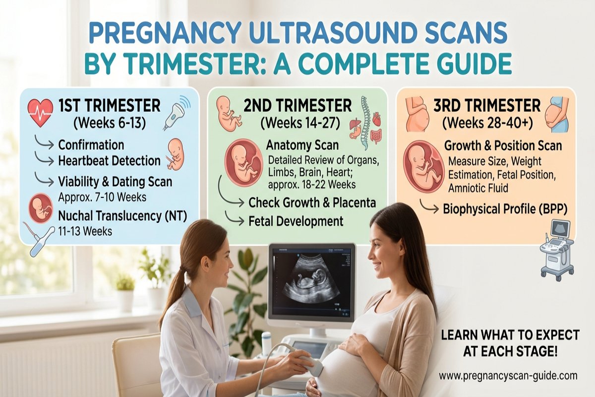
The first trimester extends from the beginning of pregnancy through 13 weeks and 6 days.
An early ultrasound may be recommended to assess the pregnancy and help establish important information about its development.
What Can an Early Pregnancy Scan Check?
Depending on the timing, an early scan may help:
Confirm that the pregnancy is located inside the uterus.
Identify the number of embryos.
Assess early pregnancy development and cardiac activity when visible.
Estimate gestational age and the expected delivery date.
Investigate symptoms such as pain or bleeding when clinically appropriate.
A scan performed very early in pregnancy may not show all expected findings. Depending on the clinical situation, a follow-up scan or blood tests may be recommended.
Nuchal Translucency and First-Trimester Screening
In some screening pathways, an ultrasound performed around 11 to 14 weeks assesses the fluid space at the back of the baby’s neck, known as nuchal translucency. This measurement may form part of a screening assessment for certain chromosomal conditions.
Nuchal translucency is a screening measurement, not a diagnosis. Other screening options, including non-invasive prenatal testing (NIPT), may be discussed according to individual circumstances and local guidance.
Second Trimester Ultrasound: The Fetal Anomaly Scan
The second trimester extends from 14 weeks through 27 weeks and 6 days.
One of the main ultrasound examinations during this period is the fetal anomaly scan, commonly performed at approximately 18 to 22 weeks, depending on local guidelines.
What Does the Fetal Anomaly Scan Assess?
The detailed scan examines fetal anatomy, including structures such as the brain, spine, heart, abdomen, kidneys, and limbs, as well as the placenta and amniotic fluid.
It may identify some structural abnormalities, but not every condition can be detected by ultrasound. The quality of the images and the findings may be affected by fetal position, gestational age, and other factors.
For a detailed explanation, read our related article: Fetal Anomaly Scan: Complete Guide to the Detailed Pregnancy Ultrasound.
Can the Scan Identify Every Birth Defect?
No. Some conditions may not be visible during the examination, may develop later, or may be difficult to detect with ultrasound. A reassuring scan is important, but it cannot guarantee that the baby has no medical condition.
If a finding needs further assessment, your healthcare professional may recommend a specialist scan, additional tests, or a fetal medicine consultation.
Third Trimester Ultrasound: Growth and Well-Being
The third trimester begins at 28 weeks and continues until delivery.
Not every pregnancy requires a routine ultrasound in the third trimester. A scan may be recommended when there is a clinical reason to assess fetal growth, placental function, amniotic fluid, or the baby’s position.
What Can a Third-Trimester Scan Assess?
Depending on the indication, the examination may evaluate:
Fetal growth: Measurements are used to estimate the baby’s size.
Amniotic fluid: The amount of fluid around the baby may be assessed.
Placental location: This may be reviewed when clinically necessary.
Fetal position: The scan can help identify whether the baby is head-down or in another position.
Doppler blood flow: This may be used when there are concerns about fetal growth or placental function.
An estimated fetal weight is not exact. It is an ultrasound-based estimate and can differ from the baby’s actual birth weight.
When Is a Growth Scan Recommended?
A growth scan may be considered if there are concerns about the baby’s size, maternal medical conditions, previous pregnancy complications, or other clinical findings.
If fetal growth restriction is suspected, the care team may recommend additional scans and Doppler assessments to guide monitoring and management.
What Is Doppler Ultrasound in Pregnancy?
Doppler ultrasound evaluates blood flow in selected blood vessels. During pregnancy, it may help assess circulation between the placenta and baby in certain clinical circumstances.
Doppler examinations can be particularly useful when a clinician is monitoring suspected fetal growth restriction or other complications that may affect placental function.
Doppler ultrasound is not required at every routine pregnancy appointment. Its use depends on the clinical indication and the healthcare professional’s assessment.
Is Ultrasound Safe During Pregnancy?
Diagnostic ultrasound has been widely used in pregnancy. When performed by trained healthcare professionals for appropriate medical purposes, it is considered a useful imaging method without ionizing radiation.
Scans should be performed when medically appropriate, using suitable equipment and exposure settings. Non-medical scans undertaken solely for keepsake images are not a substitute for clinical examinations.
If you have questions about the purpose or timing of a scan, discuss them with your obstetrician or fetal medicine specialist.
How Should You Prepare for a Pregnancy Ultrasound?
Preparation depends on the type of scan and the stage of pregnancy.
Before your appointment:
Follow the clinic’s instructions about whether you need a full bladder.
Bring relevant pregnancy records and previous scan reports if requested.
Tell the healthcare professional about important medical history or symptoms.
Ask what the examination is intended to assess and when results will be discussed.
Some early pregnancy scans may require a full bladder, while others may not. Follow the specific instructions provided by your clinic.
Frequently Asked Questions
How many ultrasound scans do I need during pregnancy?
The number varies according to local antenatal care guidelines and individual medical needs. Some pregnancies involve routine scans at specific stages, while others require additional examinations.
When is the best time for the fetal anomaly scan?
The detailed fetal anomaly scan is commonly performed around 18 to 22 weeks, although timing can vary according to local guidelines and individual circumstances.
Can ultrasound detect all fetal abnormalities?
No. Ultrasound can identify many structural abnormalities, but some conditions cannot be detected or may not be visible at the time of the scan.
Will I need a third-trimester scan?
Not everyone needs one. Your clinician may recommend a third-trimester scan if there are concerns about fetal growth, amniotic fluid, placental location, or another pregnancy-related issue.
What happens if an ultrasound finding is unclear?
Your healthcare professional may recommend a repeat scan, a more detailed examination, or a specialist consultation. An unclear finding does not automatically mean that a problem is present.
Conclusion
Pregnancy ultrasound scans provide valuable information throughout pregnancy, from early assessment to detailed fetal anatomy and growth monitoring. The purpose and timing of each scan depend on gestational age, clinical findings, and individual medical needs.
Regular antenatal care helps ensure that recommended examinations are arranged at the appropriate time and that any concerns are assessed promptly.
For personalized advice about pregnancy ultrasound, fetal growth, or specialist assessment, consult your obstetrician or fetal medicine specialist.
Medical disclaimer: This article is intended for educational purposes only and does not replace professional medical advice, diagnosis, or treatment.